teena

teena
my baby girl

Tuesday 5 June 2012

another site dissection frog


Frog Dissection


Need a frog dissection worksheet?  Try External Anatomy | Digestive & Urogenital | Brain & Bones
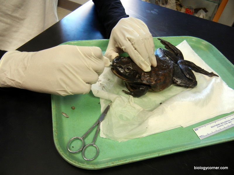
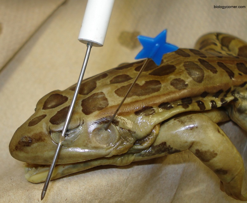
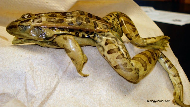
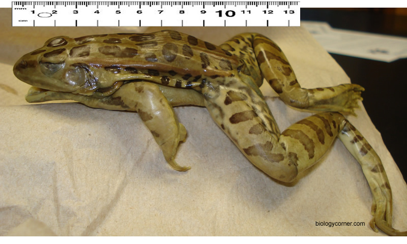
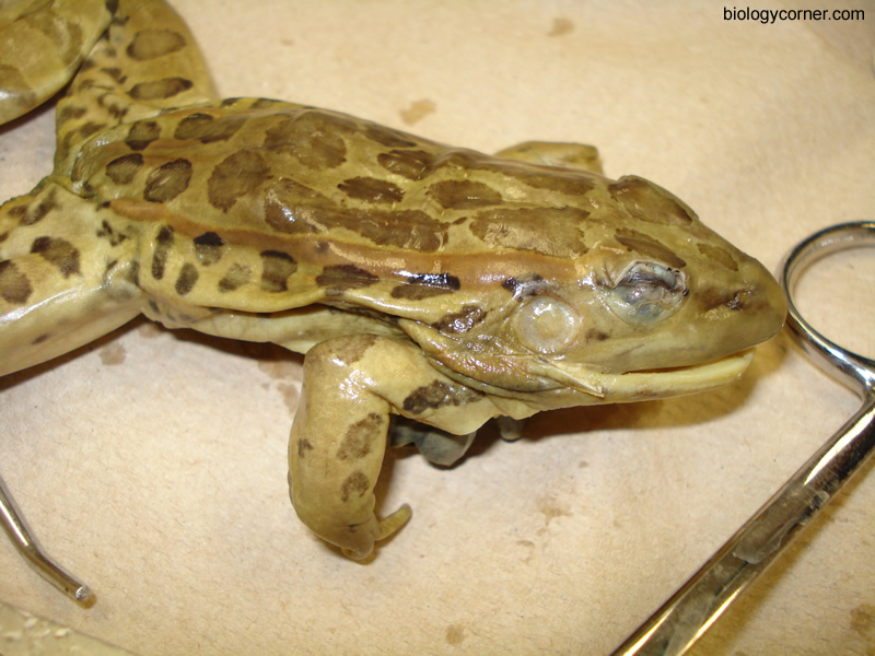
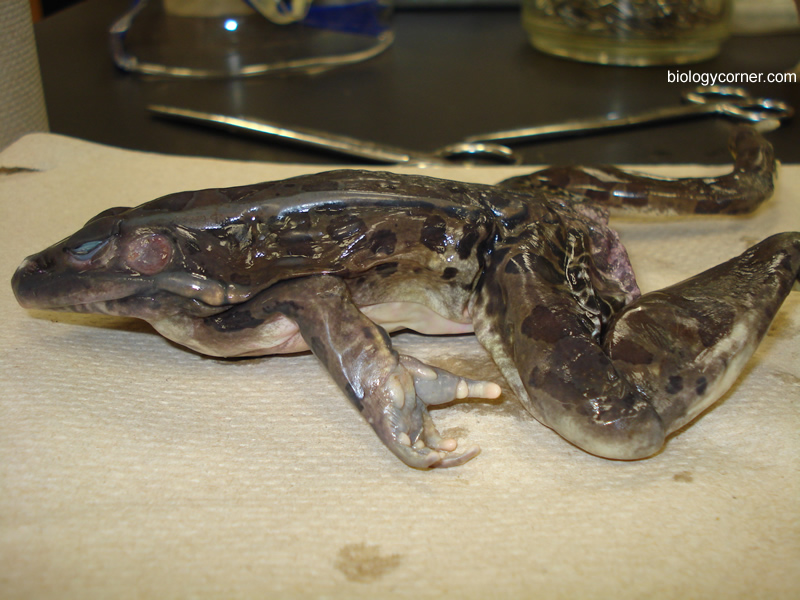
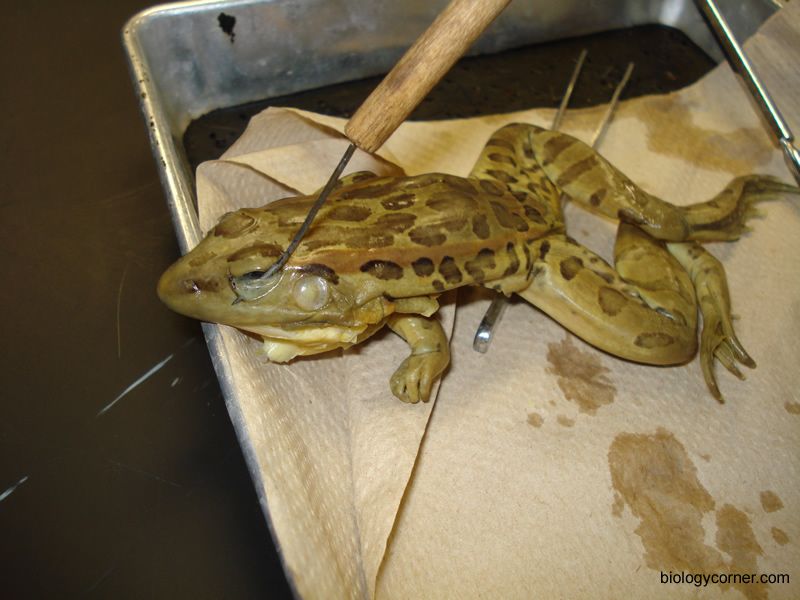
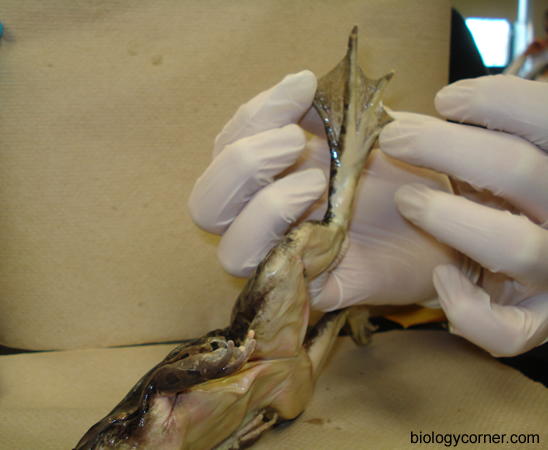
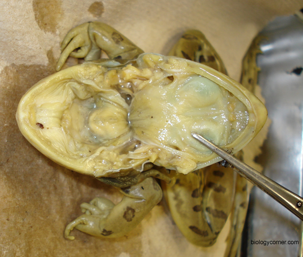
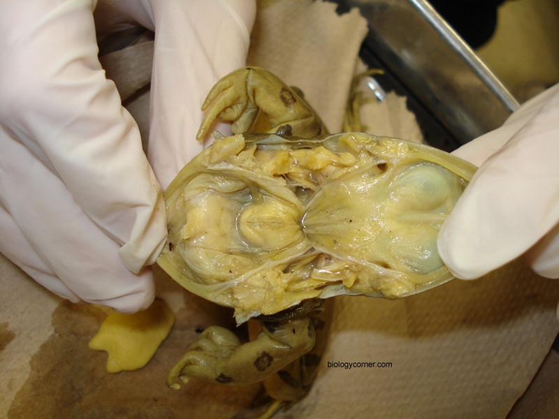
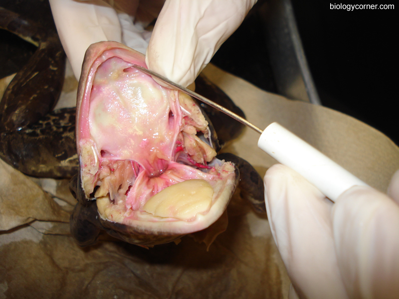
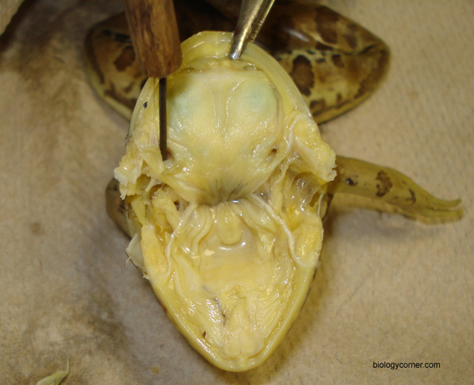
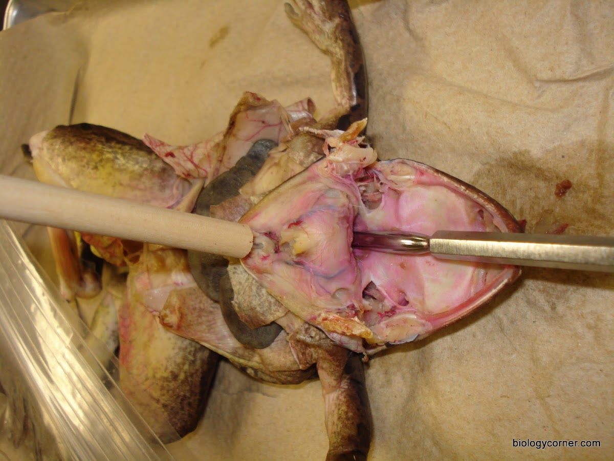
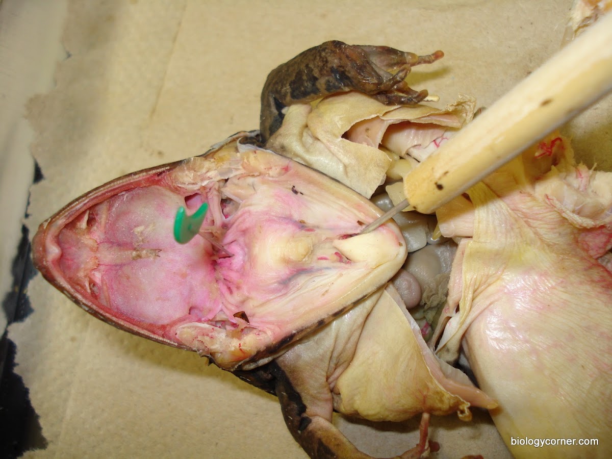
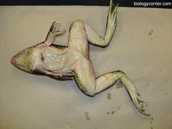
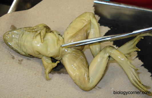
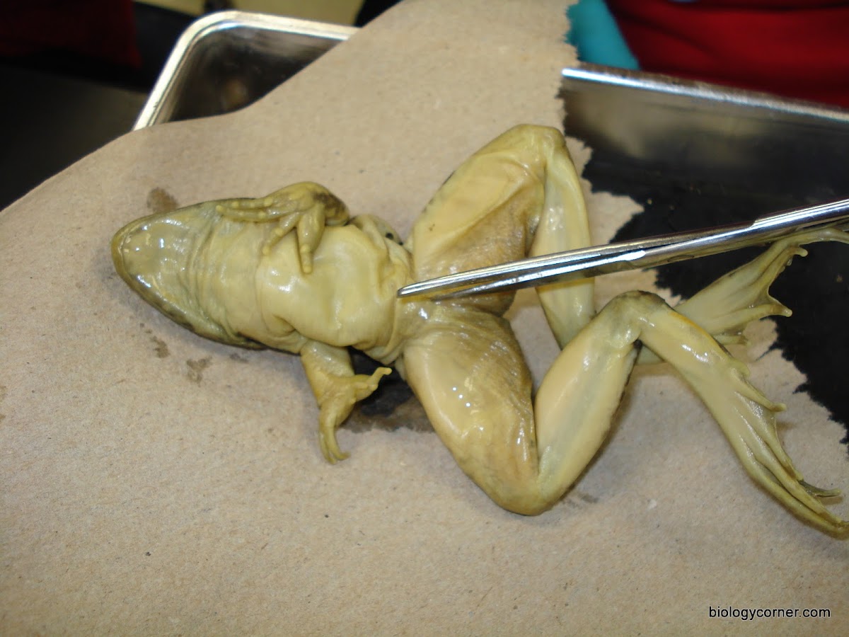
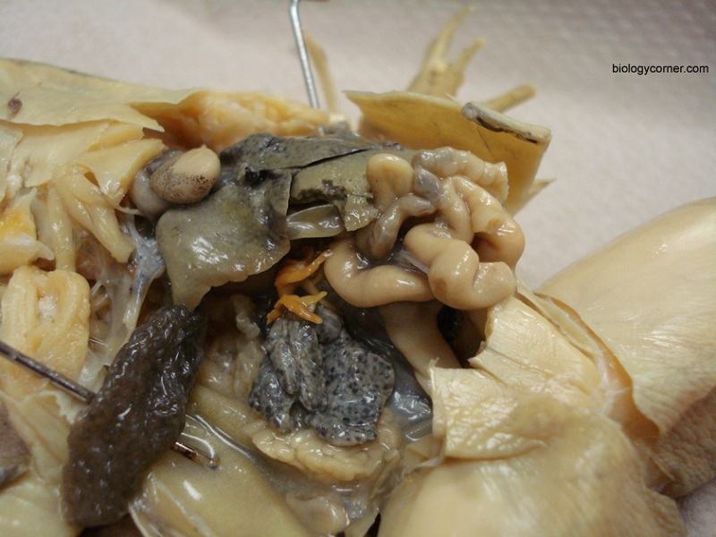
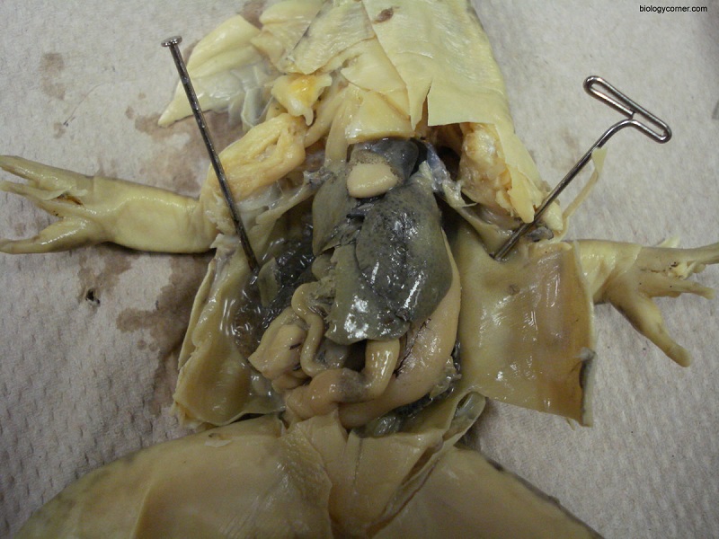
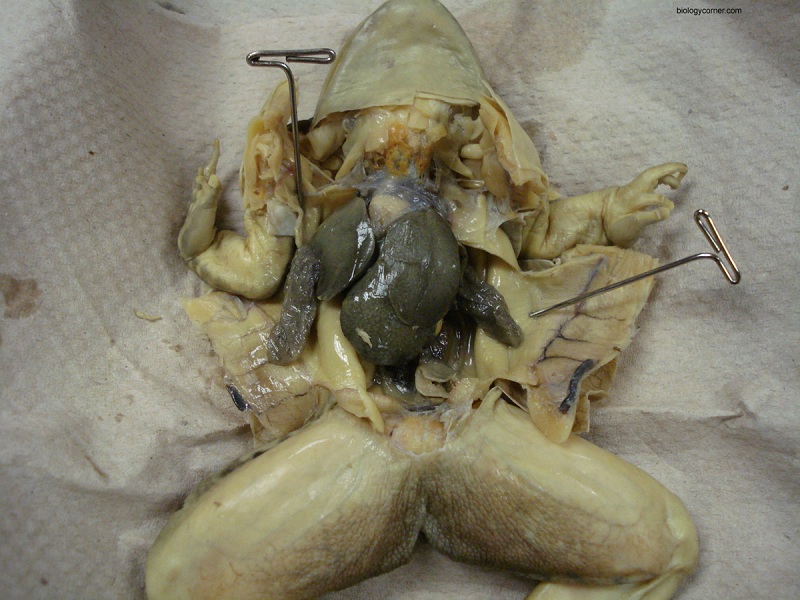
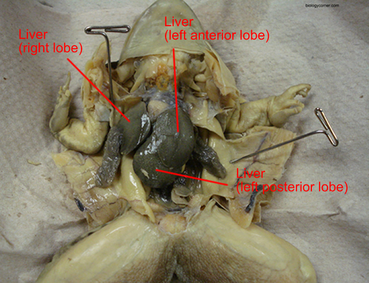
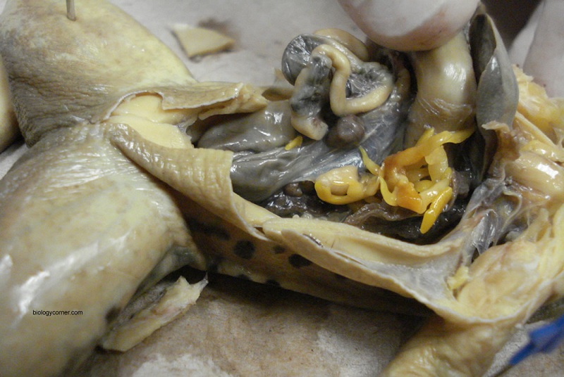
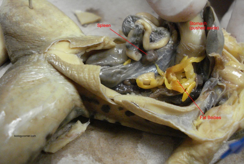
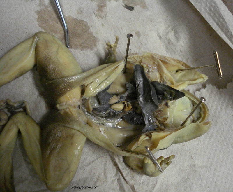
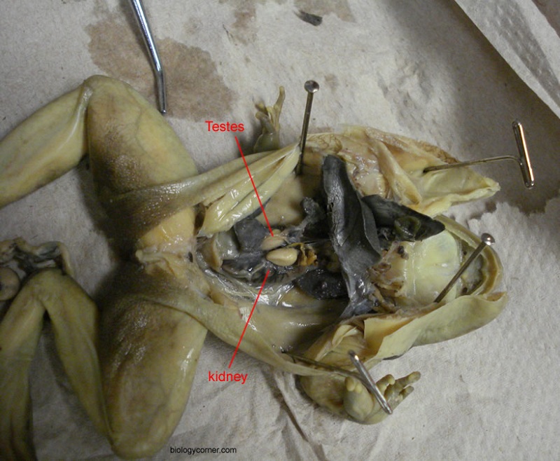
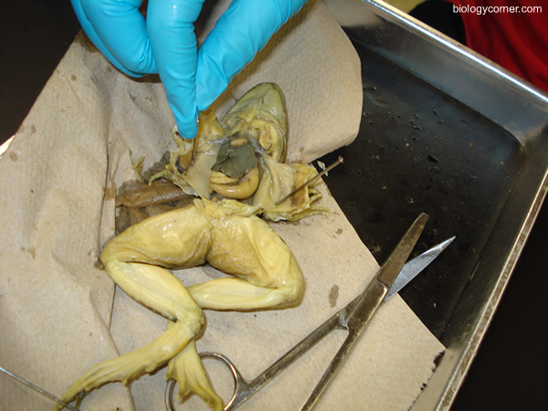
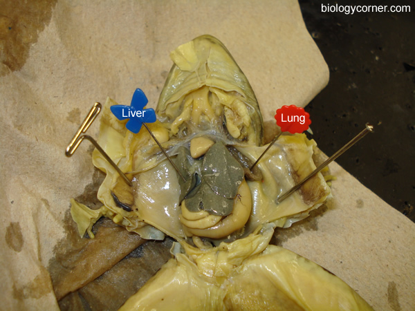
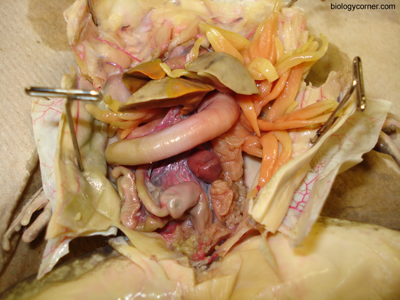
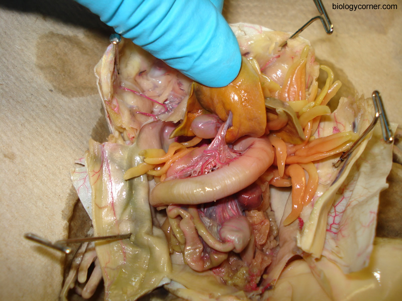
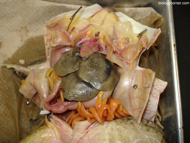
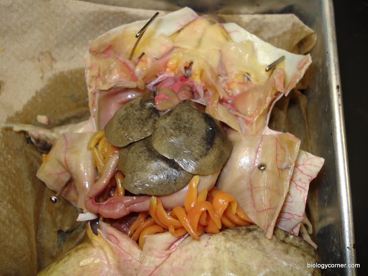
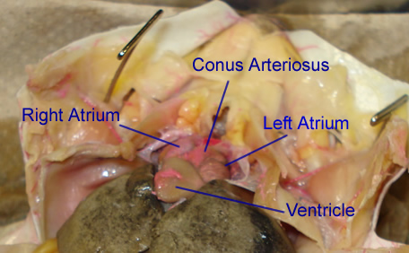
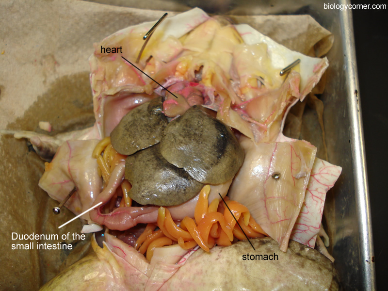
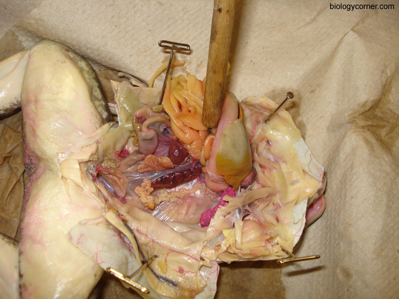
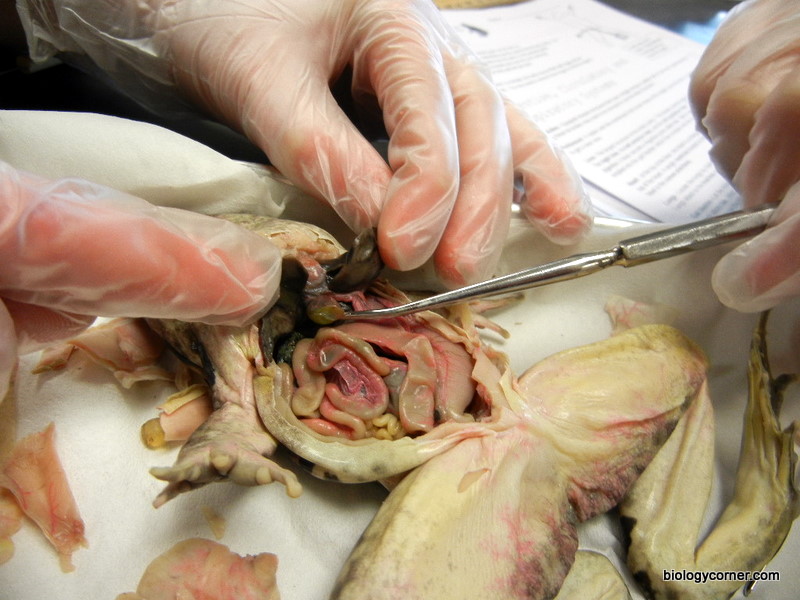
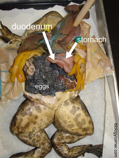
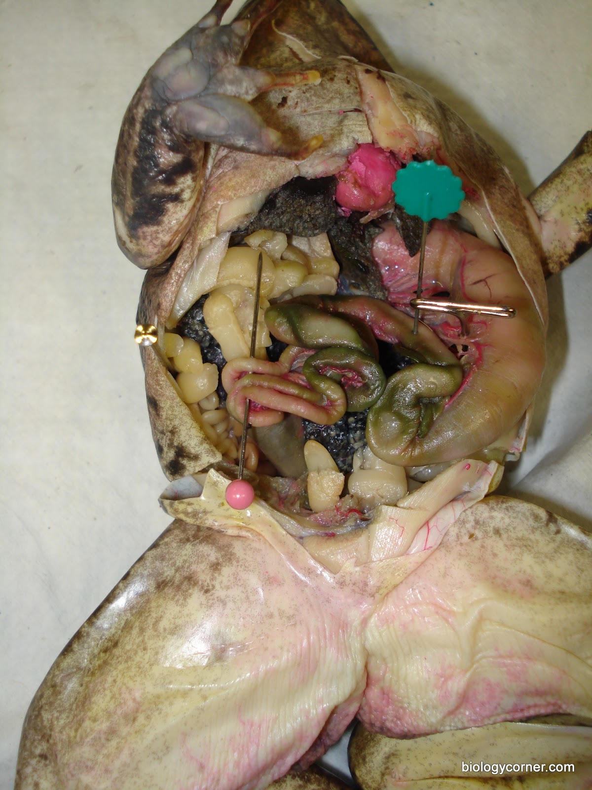
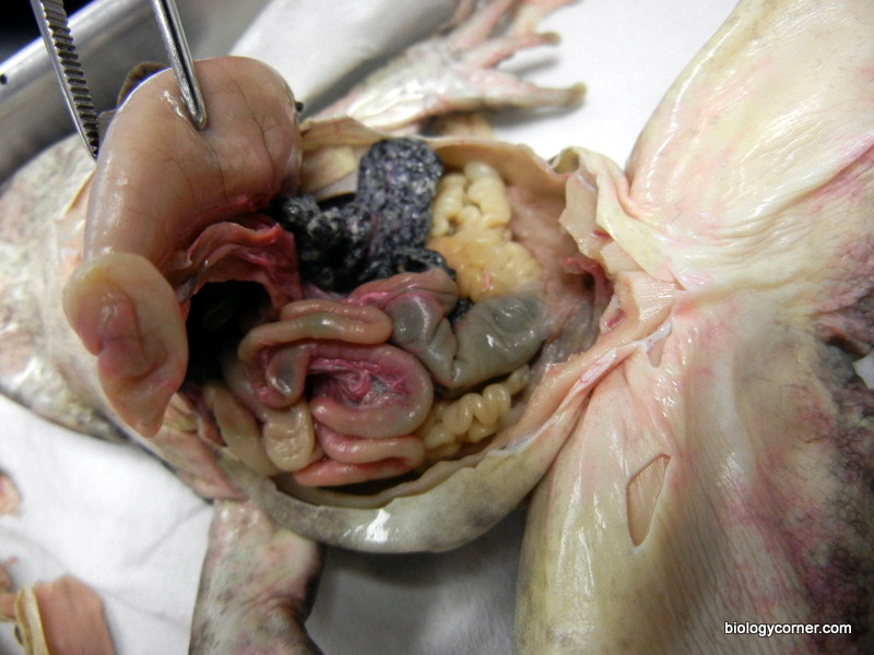
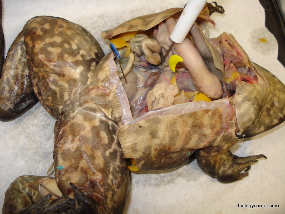
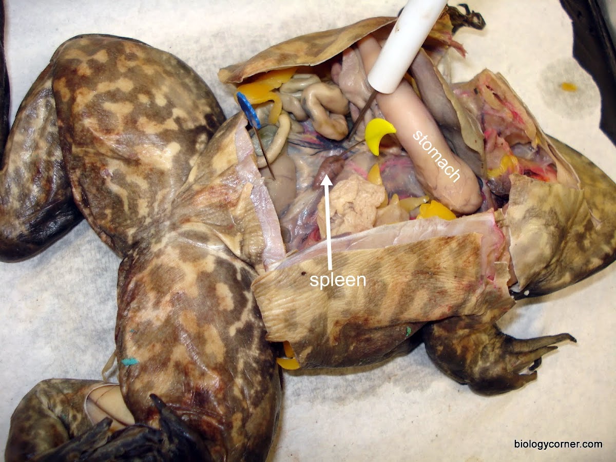
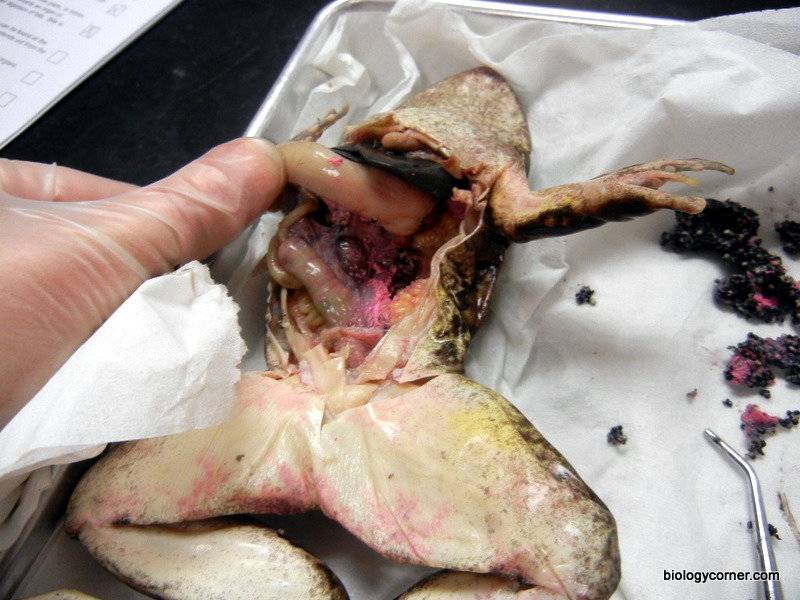
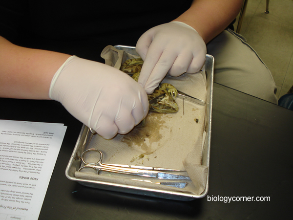
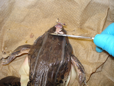
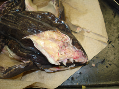
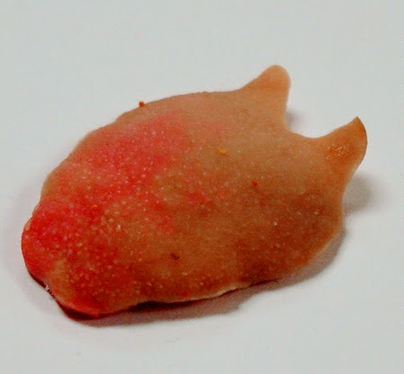
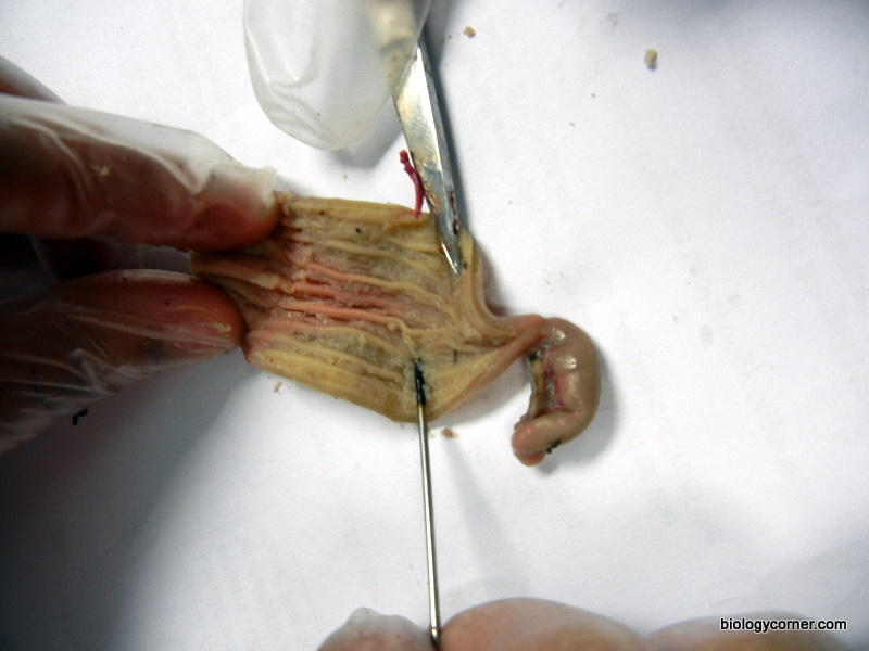
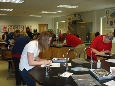
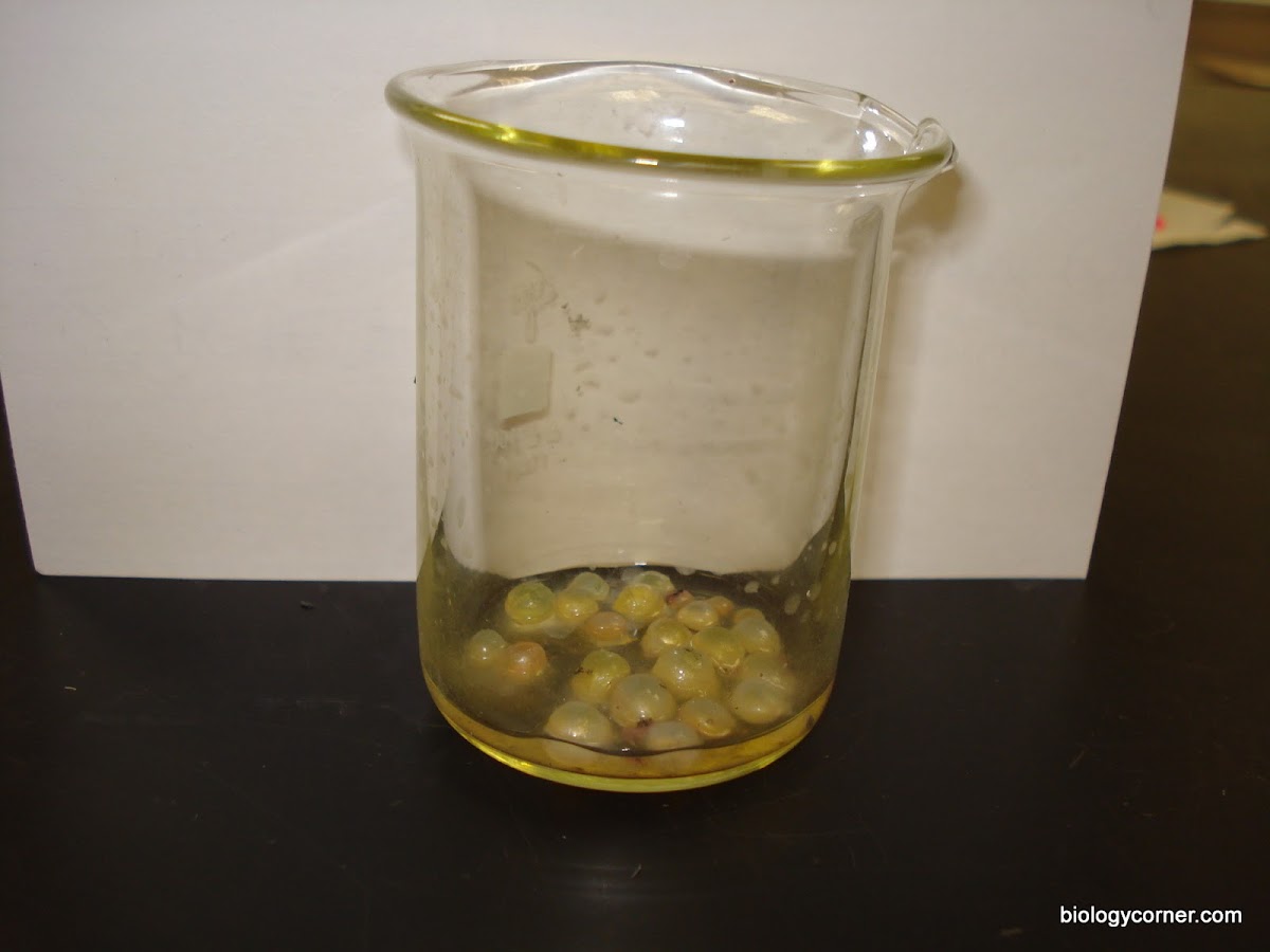
The following photos show all of the structures that are visible during a frog dissection, such as the liver, gall bladder, stomach, intestine, spleen, eggs.   Many of the photos are labeled, though some are not labeled so that they can be used in slide tests.
bullfrog.JPGdissect05.jpgdissect01.jpgdissect04.jpgdissect04_labeled.jpgdissect04_ruler.jpgdissect14.jpgdissect18_foreleg.jpgdissect15.jpgdissect19_hindleg.jpgdissect02.jpgdissect02_labeled.jpgdissect03.jpgdissect21.jpgdissect09.jpgeustachian_tubes.JPGeustachian_tubes.JPGmouth1.JPGmouth2.JPGmouth3.JPGdissect17_ventral.jpgdissect20_procedure.jpgfirst_incision.JPGdissect07.jpgdissect08.jpgdissect08_labeled.jpgdissect10.jpgdissect10_labeled.jpgdissect11.jpgdissect11_labeled2.jpgdissect12.jpgdissect12_labeled.jpgdissect13_male.jpgdissect13_male_labeled.jpgdissect22_pinning.jpgdissect23.jpgdissect23_labeled.jpgdissect24.jpgdissect25.jpgdissect26.jpgliver.JPGdissect26_heart_labeled.jpgdissect26_labeled.jpgdissect27.jpgdissect27_labeled.jpgfrog_gall_bladder.JPGfrog_liver_eggs.JPGdissect28.jpgdissect29_eggs.jpgdissect29_eggs.jpgdissect31.jpgfrog_duodenum.JPGfrog_duodenum.JPGfrog_stomach_lifted.JPGfrog_spleen.JPGfrog_spleen.JPGfrog_spleen_revealed.JPGfrog_liver_heart.JPGdissect_procedure.jpgdissect32_brain.jpgdissect33_brain.jpgfrog_tongue.JPGtongue.jpgstomach_rugae.JPGlab_practical.jpgfrog_brain.JPGdissect16_leg.jpgfrog_eyes.JPG
Please credit biologycorner.com whenever a resource is used. If resources from this site are incorporated into a website, please include link to www.biologycorner.com

hotchalk logo

No comments:

Post a Comment